Computed Tomography (CT) Scan of the Spine
Test Overview
A CT scan uses X-rays to make detailed pictures of the spine and vertebrae.
During the test, you will lie on a table that is attached to the CT scanner, which is a large doughnut-shaped machine. The CT scanner sends X-rays through the body. Each rotation of the scanner takes a second and provides a picture of a thin slice of the organ or area being studied. One part of the scanning machine can tilt to follow the curve of your spine. All of the pictures are saved as a group on a computer. They also can be printed.
In some cases, a dye called contrast material may be put in a vein (IV) in your arm or into the spinal canal. The dye makes structures and organs easier to see on the CT pictures. The dye may be used to check blood flow and look for tumors, areas of inflammation, or nerve damage.
Why It Is Done
A CT scan of the spine is done to:
- Look at the bones of the spine (vertebrae).
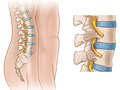
- Find problems of the spine, such as tumors, fractures, deformities, infection, or narrowing of the spinal canal (spinal stenosis).
- Find a herniated disc of the spine.
- Check to see if osteoporosis has caused compression fractures.
- Check on problems of the spine that have been present since birth (congenital).
- Look at problems seen during a standard X-ray test.
- Check how well spinal surgery or therapy is working for a spine problem.
How To Prepare
Before the CT scan, tell your doctor if you:
- Are or might be pregnant.
- Are allergic to any medicines, including iodine dyes.
- Have a heart condition, such as heart failure.
- Have diabetes.
- Take metformin. You may have to adjust your medicine for a day before and after the test.
- Have had kidney problems.
- Have asthma.
- Have had multiple myeloma.
- Have had an X-ray test using barium contrast material (such as a barium enema) in the past 4 days. Barium shows up on X-ray films and makes it hard to see the picture clearly.
- Become very nervous in small spaces. You need to lie still inside the CT scanner, so you may need a sedative to help you relax.
Arrange for someone to take you home in case you get a sedative for the test.
Talk to your doctor about any concerns you have regarding the need for the test, its risks, or how it will be done. To help you understand the importance of this test, fill out the medical test information form( What is a PDF document? ).
How It Is Done
A CT scan is usually done by a radiology technologist. The pictures are usually read by a doctor (radiologist). Other doctors also may review a CT scan.
You may need to take off any jewelry. You will need to take off all or most of your clothes, depending on which area is studied. You may be able to wear your underwear for some scans. You will be given a gown to use during the test.
During the test, you will lie on a table that is attached to the CT scanner.
The table slides into the round opening of the scanner, and the scanner moves around your body. The table will move while the scanner takes pictures. You may hear a click or buzz as the table and scanner move. It is very important to lie still during the test.
During the test, you may be alone in the scanning room. But the technologist will watch you through a window. You will be able to talk to the technologist through a two-way intercom.
The test will take about 30 to 60 minutes. Most of this time is spent getting ready for the scan. The actual scan only takes a few seconds.
CT scan with contrast (CT myelogram)
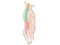
A standard CT scan may be done before the contrast material for a CT myelogram is given. The dye is usually put in the space around your spinal cord. A sample of the fluid from the spinal canal (cerebrospinal fluid) may be taken out so other tests can be done on it.
If dye is placed in your back, you will lie on your stomach or on your side on a table. The dye is usually put in your lower back but may be put in at the base of your skull. The skin over the site may be shaved. It will be cleaned. The area around the site may be numbed with medicine.
The table may be tilted or you may be moved into different positions so the dye moves to different areas of the spine.
You need to lie very still so the dye stays in the right place for clear pictures. Your pulse, breathing rate, and blood pressure may be checked during the test.
In some cases, the dye can also be put in a vein (IV) in your arm.
A CT scan with contrast material usually takes 15 to 30 minutes. Drink lots of liquids for 24 hours after the scan to help flush the dye out of your body.
How It Feels
The test will not cause pain. The table you lie on may feel hard, and the room may be cool. It may be hard to lie still during the test.
Some people feel nervous inside the CT scanner.
If you get medicine to help you relax or if contrast material is used, you may have an IV put in your hand or arm. You may feel a quick sting or pinch when the IV is started. The dye may make you feel warm and flushed and give you a metallic taste in your mouth. Some people feel sick to their stomach or get a headache. Tell the technologist or your doctor how you are feeling.
If you have dye put in your back, you may feel a sting or pinch when the needle is put in.
After a test in which the dye is put in your back, you will be told to keep your head up and to not bend over or lie flat. This will help prevent headaches and seizures.
Risks
The chance of a CT scan causing a problem is small.
- There is a chance of an allergic reaction to the contrast material.
- If you breastfeed and are concerned about whether the dye used in this test is safe, talk to your doctor. Most experts believe that very little dye passes into breast milk and even less is passed on to the baby. But if you prefer, you can store some of your breast milk ahead of time and use it for a day or two after the test.
- If you have diabetes or take metformin (Glucophage), the dye may cause problems. Your doctor will tell you when to stop taking metformin and when to start taking it again after the test so you will not have problems.
- You may have nausea or vomiting after the test.
- There is a small chance of an infection at the needle site on your spine or bleeding into the space around the spinal cord.
- An injection into the space around the spinal cord may cause a headache. In rare cases, seizures may occur after an injection of contrast material.
- There is a small chance of getting cancer from some types of CT scans.footnote 1 The risk is higher in children, young adults, and people who have many radiation tests. If you are concerned about this risk, talk to your doctor about the benefits and risks of a CT scan, and confirm that the test is needed.
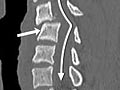
Results
A computed tomography (CT) scan uses X-rays to make detailed pictures of the spine and vertebrae in the neck (cervical spine), upper back (thoracic spine), or lower back (lumbosacral spine).
Complete results usually are ready for your doctor in 1 to 2 days.
|
Normal: |
Spinal bones (vertebrae) are normal in shape, number, and alignment. |
|
The discs and joints that support the spine are normal. |
|
|
The spinal canal is normal in size and shape. |
|
|
If contrast material is used, it flows evenly through the spinal canal. No narrowing or blockage of the spinal canal is present. |
|
|
None of the nerves leaving the spinal cord are compressed or pinched. No growths or bulges are present. |
|
|
Abnormal: |
Spinal bones (vertebrae) are missing, damaged, or out of alignment. |
|
One or more discs may be damaged. One or more herniated discs are found. |
|
|
The flow of contrast material through the spinal canal is restricted or blocked, indicating narrowing of the canal (spinal stenosis). |
|
|
The vertebrae show signs of arthritis or bone problems caused by osteoporosis. |
|
|
A condition that has been present from birth (congenital condition) is present in the spine or the vertebrae. |
|
|
An abscess or spinal tumor is found. |
What Affects the Test
The following may stop you from having the test or may change the test results:
- Pregnancy. CT scans are not usually done during pregnancy.
- Barium used for another test. Barium shows up on a CT scan. If a CT scan of the lower back is needed, it should be done before any tests that use barium, such as a barium enema.
- Metal objects in the body. These items, such as surgical clips or metal in joint replacements, may prevent a clear view of the body area.
- You are not able to lie still during the test.
What To Think About
- Sometimes your CT test results may be different than those from other types of X-ray tests, magnetic resonance imaging (MRI), or ultrasound scans, because the CT scan provides a different view.
- Children who need a CT scan may need special instructions for the test. If the child is too young to hold still or is afraid, the doctor may give the child a medicine (sedative) to help him or her relax.
- If your child is scheduled for a CT scan, talk with your child’s doctor about the need for the scan and the risk of radiation exposure to your child.
- CT results are often compared to positron emission tomography (PET) results to help find cancer. Some new scanners do both scans at the same time.
- MRI may give more information than a CT scan about the spinal discs and spinal cord. To learn more, see the topic Magnetic Resonance Imaging (MRI).
- When a CT scan of the spine is done with a myelogram, it is called a CT myelogram. An MRI of the spine is often done in place of a CT myelogram. To learn more, see the topic Myelogram.
References
Citations
- Einstein AJ, et al. (2007). Estimating risk of cancer associated with radiation exposure from 64-slice computed tomography coronary angiography. JAMA, 298(3): 317–323.
Other Works Consulted
- Fischbach FT, Dunning MB III, eds. (2009). Manual of Laboratory and Diagnostic Tests, 8th ed. Philadelphia: Lippincott Williams and Wilkins.
- Pagana KD, Pagana TJ (2010). Mosby’s Manual of Diagnostic and Laboratory Tests, 4th ed. St. Louis: Mosby.
- Pearce MS, et al. (2012). Radiation exposure from CT scans in childhood and subsequent risk of leukaemia and brain tumours: A retrospective cohort study. Lancet, 380(9840): 499–505.
- U.S. Food and Drug Administration (2008). FDA preliminary public health notification: Possible malfunction of electronic medical devices caused by computed tomography (CT) scanning. Available online: http://www.fda.gov/MedicalDevices/Safety/AlertsandNotices/PublicHealthNotifications/ucm061994.htm.
Current as of: March 28, 2019
Author: Healthwise Staff
Medical Review:Adam Husney, MD – Family Medicine & E. Gregory Thompson, MD – Internal Medicine & Martin J. Gabica, MD – Family Medicine & Howard B. Schaff, MD – Diagnostic Radiology
This information does not replace the advice of a doctor. Healthwise, Incorporated, disclaims any warranty or liability for your use of this information. Your use of this information means that you agree to the Terms of Use. Learn how we develop our content.