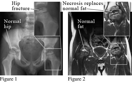
X-Ray and MRI of Legg-Calve-Perthes Disease
Courtesy of Paul Traughber, M.D., Boise, Idaho.
Figure 1 is an X-ray of a child’s normal hipbone and a broken (fractured) hipbone from poor blood flow because of Legg-Calve-Perthes disease (LCPD). Figure 2 is an MRI of a child’s normal hipbone with fat in the growth center and an abnormal hipbone where the fat has been lost because of LCPD.
Current as of: June 26, 2019
Author: Healthwise Staff
Medical Review:William H. Blahd, Jr., MD, FACEP – Emergency Medicine & Adam Husney, MD – Family Medicine & Kathleen Romito, MD – Family Medicine & Kenneth J. Koval, MD – Orthopedic Surgery, Orthopedic Trauma