Cystourethrogram
Test Overview
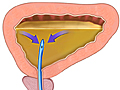
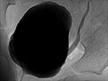
A cystourethrogram is an X-ray test that takes pictures of your bladder and urethra while your bladder is full and while you are urinating. A thin flexible tube (urinary catheter) is inserted through your urethra into your bladder. A liquid material that shows up well on an X-ray picture (contrast material) is injected into your bladder through the catheter, then X-rays are taken with the contrast material in your bladder. More X-rays may be taken while urine flows out of your bladder, in which case the test is called a voiding cystourethrogram (VCUG).
If X-rays are taken while contrast material is being injected into the urethra, the test is called a retrograde cystourethrogram because the contrast material flows into the bladder opposite the usual direction of urine flow.
Why It Is Done
A cystourethrogram is done to:
- Find the cause of repeated urinary tract infections.
- Look for injuries to the bladder or urethra.
- Find the cause of urinary incontinence.
- Check for structural problems of the bladder and urethra.
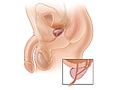
- Look for enlargement (hypertrophy) of the prostate or narrowing (stricture) of the urethra in men.
- Find out if urine is flowing the wrong way, from the bladder back toward the kidneys (vesicoureteral reflux).
- Look more carefully at abnormalities first found by intravenous pyelography.
How To Prepare
Tell your doctor if:
- You are or might be pregnant.
- You have symptoms of a urinary tract infection, such as pain or burning when you urinate.
- You are allergic to the iodine dye used in the contrast material or any other substance that contains iodine. Also tell your doctor if you are allergic to any medicines or have ever had a serious allergic reaction (anaphylaxis), such as after being stung by a bee or from eating shellfish.
- Within the past 4 days, you have had an X-ray test using barium contrast material, such as a barium enema, or have taken a medicine (such as Pepto-Bismol) that contains bismuth. Barium and bismuth can interfere with test results.
- You have an intrauterine device (IUD) in place.
This test is often done in children to see if they may have an abnormal backflow of urine (vesicoureteral reflux). Prepare your child for exams and tests that are needed by explaining them in a simple way. Use positive words as much as possible. Doing so will help your child understand what to expect and can help reduce fears.
You may be asked to sign a consent form that says you understand the risks of the test and agree to have it done.
Talk to your doctor about any concerns you have regarding the need for the test, its risks, how it will be done, or what the results may mean. To help you understand the importance of this test, fill out the medical test information form( What is a PDF document? ).
How It Is Done
A cystourethrogram is done by a urologist or a radiologist. The doctor may be assisted by an X-ray technologist. You usually will not have to be admitted to the hospital.
You will need to take off all or most of your clothes, and you will be given a cloth or paper covering to use during the test. You will be asked to urinate just before the test begins.
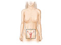
You will be asked to lie on your back on an X-ray table. Your genital area will be cleaned and draped with sterile towels. Men may be given a lead shield that covers their genitals to protect them from radiation. But women’s ovaries cannot be shielded without blocking the view of the bladder.
A catheter will be placed through your urethra and into your bladder. Contrast material will then slowly be injected through the catheter until your bladder is full.
X-rays will be taken when you are standing up and sitting and lying down. The catheter is removed and more X-rays will be taken while you are urinating. You may be asked to stop urinating, change positions, and begin urinating again. If you are unable to urinate in one position, you may be asked to try it from another position.
This test usually takes 30 to 45 minutes.
How It Feels
You will feel no discomfort from the X-rays. The X-ray table may feel hard and the room may be cool. You may find that the positions you need to hold are uncomfortable or painful.
You will feel a strong urge to urinate at times during the test. You may also find it somewhat uncomfortable when the catheter is inserted and left in place. You will have a feeling of fullness in your bladder and an urge to urinate when the contrast material is injected. You may be sore afterward. If so, soaking in a warm tub bath may help.
You may feel embarrassed at having to urinate in front of other people. This procedure is quite routine for the X-ray staff. If you find yourself feeling embarrassed, take deep, slow breaths and try to relax.
You may feel burning when you urinate.
Risks
A cystourethrogram does not usually cause problems. Occasionally this test may lead to a urinary tract infection. If the contrast material is injected with too much pressure, there is some chance of damage to the bladder or urethra.
There is always a slight chance of damage to cells or tissue from radiation, including the low levels of radiation used for this test. But the chance of damage from the X-rays is usually very low compared with the benefits of the test.
Some people may have an allergic reaction to the contrast material.
After the test
After the test, you may need to urinate frequently, with some burning during and after urination for a day or two. Drink lots of fluids to help minimize the burning and to prevent a urinary tract infection.
A pinkish tinge to the urine is common for several days after a cystourethrogram. But call your doctor immediately if:
- Your urine remains red or you see blood clots after you have urinated several times.
- You have not been able to urinate 8 hours after the test.
- You have a fever, chills, or severe pain in your flank or abdomen. These may be signs of a kidney infection.
- You have symptoms of a urinary tract infection (UTI). These symptoms include:
- Pain or burning upon urination.
- An urge to urinate frequently, but usually passing only small quantities of urine.
- Dribbling or leakage of urine.
- Urine that is reddish or pinkish, foul-smelling, or cloudy.
- Pain or a feeling of heaviness in the lower abdomen.
Results
A cystourethrogram is an X-ray test that takes pictures of your bladder and urethra while you are urinating. Some results may be available immediately after the cystourethrogram. Final results are usually available within 1 to 2 days.
|
Normal: |
The bladder appears normal. Urine flows normally from the bladder. The bladder empties all the way. The contrast material flows evenly out of the bladder through a smooth-walled urethra. |
|
Abnormal: |
Bladder stones, tumors, narrowing or pouches in the wall (diverticula) of the urethra or bladder are seen in the bladder. If the test was done because of possible injury to the bladder, a tear is found in the bladder wall or urethra. Urine flows backward from the bladder into the ureters (vesicoureteral reflux). Contrast material leaks from the bladder. The bladder does not empty all the way. The prostate gland is enlarged. |
What Affects the Test
Reasons you may not be able to have the test or why the results may not be helpful include:
- Having barium (from a previous barium enema test), gas, or stool in the bowel.
- Being unable to urinate on command.
- Pain caused by having the catheter in the urethra. This may also cause problems with your urinary stream. You may have a muscle spasm or not be able to fully relax the muscles that control your bladder.
A cystourethrogram is not usually done during pregnancy because the X-rays could harm an unborn baby.
What To Think About
Other tests that use X-rays and contrast material to look for problems in the kidney, bladder, and urethra include:
- Retrograde urethrogram. This test is sometimes used to check for problems with a man’s urethra. A small quantity of contrast material is injected into the urethra through a catheter. X-rays are then taken. This test can help find tears, scar tissue, prostate narrowing, tumors, or malformations of the urethra.
- Whitaker test. Also called a pressure/flow study, this test combines X-rays with measurements of pressure and flow in the kidneys and ureters. It is used to find the cause of blockage of the kidneys.
- Intravenous pyelogram (IVP) IVP is commonly done to diagnose certain diseases of the urinary tract (such as kidney stones, tumors, or infection) and detect abnormalities of the urinary tract that were present from birth (congenital). It can show the size, shape, and position of the kidneys, the bladder, the ureters, and the urethra, and it can evaluate the collecting system inside the kidneys. To learn more, see the topic Intravenous Pyelogram (IVP).
References
Other Works Consulted
- Chernecky CC, Berger BJ (2008). Laboratory Tests and Diagnostic Procedures, 5th ed. St. Louis: Saunders.
- Fischbach FT, Dunning MB III, eds. (2009). Manual of Laboratory and Diagnostic Tests, 8th ed. Philadelphia: Lippincott Williams and Wilkins.
- Pagana KD, Pagana TJ (2010). Mosby’s Manual of Diagnostic and Laboratory Tests, 4th ed. St. Louis: Mosby Elsevier.
Current as of: December 19, 2018
Author: Healthwise Staff
Medical Review:E. Gregory Thompson, MD – Internal Medicine & Adam Husney, MD – Family Medicine & Avery L. Seifert, MD, FACS – Urology

This information does not replace the advice of a doctor. Healthwise, Incorporated, disclaims any warranty or liability for your use of this information. Your use of this information means that you agree to the Terms of Use. Learn how we develop our content.