Encephalitis
Current as of: June 9, 2019
Author: Healthwise Staff
Medical Review:Kathleen Romito, MD – Family Medicine & Adam Husney, MD – Family Medicine
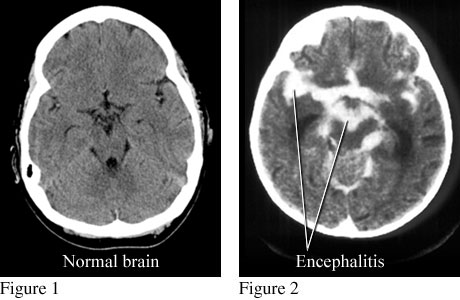
Courtesy of Paul Traughber, M.D., Boise, Idaho. Figure 1 is a computed tomography (CT) scan of a normal brain. Figure 2 is a CT scan that shows an accumulation of contrast material in infected areas and around the brain from encephalitis.
Current as of: June 9, 2019
Author: Healthwise Staff
Medical Review:Kathleen Romito, MD – Family Medicine & Adam Husney, MD – Family Medicine