Abdominal Ultrasound
Test Overview
An abdominal ultrasound takes pictures of the organs and other structures in your upper belly. It uses sound waves to show images on a screen.
Areas that can be checked include the:
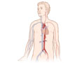
- Abdominal aorta. This large blood vessel passes down the back of the chest and belly. It supplies blood to the lower part of the body and the legs.
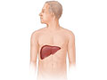
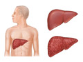
- Liver. This large organ lies under the rib cage on the right side of the belly. It makes bile (a substance that helps digest fat). It also stores sugars and breaks down many of the body’s waste products.
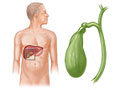
- Gallbladder. This small organ is right under the liver. It stores bile. When you eat, the gallbladder contracts to send bile to the bowels (intestines). The bile helps your body digest food and absorb vitamins that dissolve in fat.
- Spleen. This organ helps fight infection. It also filters old red blood cells. The spleen sits to the left of the stomach, just behind the lower left ribs.
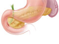
- Pancreas. This gland is in the upper belly. It makes enzymes that help digest food. The digestive enzymes then move into the bowels. The pancreas also releases insulin into the bloodstream. Insulin helps the body use sugars for energy.
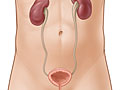
- Kidneys. This pair of organs is in the upper part of your belly, but toward your back. The kidneys remove wastes from the blood. They also make urine.
If your doctor needs more details about a specific organ in the upper part of your belly, you may get a special ultrasound, such as a kidney ultrasound. If you need the structures and organs in your lower belly checked, you will get a pelvic ultrasound.
Why It Is Done
Abdominal ultrasound is done to:
- Find the cause of belly pain.
- Find, measure, or monitor an aneurysm in the aorta. An aneurysm may cause a large, pulsing lump in the belly.
- Check the size, shape, and position of the liver. It may also check for problems of the liver. These include jaundice, cirrhosis, or fatty liver. This test may be done to follow up after liver function tests.
- Look for gallstones, inflammation of the gallbladder, or blocked bile ducts.
- Learn the size of an enlarged spleen and look for damage or disease.
- Find problems with the pancreas, such as a tumor.
- Look for blocked urine flow in a kidney. If needed, a kidney ultrasound can find out the size of the kidneys, detect a mass, or detect fluid surrounding the kidneys. It can also look for causes of bladder infections that won’t go away. Or it can check how the kidneys are doing after a transplant.
- Find out if a mass in a belly organ is a solid tumor or a fluid-filled cyst.
- Guide the placement of a needle or other tool during a biopsy.
- Look for fluid buildup in the belly cavity. This problem is called ascites. An ultrasound also may be done to guide the needle during a paracentesis. This is a procedure to remove fluid from the belly cavity.
How To Prepare
Tell your doctor if you have had a barium enema or upper GI (gastrointestinal) tests within the past 2 days. Barium that stays in the intestines can affect the results of the ultrasound.
You may need to prepare in other ways too. It depends on what test you are having. For example:
- If you are having your liver, gallbladder, spleen, and pancreas checked, you may need to eat a fat-free meal on the evening before the test. Then you may need to avoid eating for 8 to 12 hours before the test.
- For a test of the kidneys, you may be asked to drink 4 to 6 glasses of liquid about an hour before the test. This is to fill your bladder. You may need to avoid eating for 8 to 12 hours before the test to avoid gas buildup in the intestines. Gas could affect the results of the kidney ultrasound.
- If you are having your aorta checked, you may need to avoid eating for 8 to 12 hours before the test.
How It Is Done
The test is done by a radiologist. This is a doctor who has special training to perform and interpret imaging tests. Sometimes an ultrasound technologist (sonographer) will do the test. In that case, a radiologist will supervise. The test is done in an ultrasound room in a hospital or doctor’s office.
You may need to take off your jewelry. You may also need to take off all or most of your clothes. It depends on which area is being examined. You will be given a cloth or paper to cover yourself during the test.
During the test
You will lie on your back (or on your side) on a padded exam table. Warmed gel will be spread on your belly or back to help the sound waves work best. A small handheld device (transducer) is pressed against your belly.
You may be asked to change positions so more scans can be done. For a kidney ultrasound, you may be asked to lie on your stomach.
You need to lie very still while the test is being done. You may be asked to take a breath and hold it for several seconds during the test. This lets the person doing the test see organs and structures more clearly.
The test usually takes 30 to 60 minutes.
After the test
You may be asked to wait until the radiologist has reviewed the images. He or she may want to take more views of some areas of your belly.
How It Feels
The gel may feel cold when it is put on your skin. But the gel may be warmed to body temperature first. You will feel light pressure from the transducer as it passes over your belly. Most people do not feel pain during the test. But if the test is being done to check damage from a recent injury, the slight pressure of the transducer may be somewhat painful. You will not hear or feel the sound waves.
Results
An abdominal ultrasound takes pictures of the organs and other structures in your upper belly. It uses sound waves to show images on a screen.
|
Normal: |
The organs have a normal size, shape, and texture. No abnormal growths are seen. No fluid is in the belly. |
|
The aorta looks normal. No aneurysms are seen. |
|
|
The thickness of the gallbladder wall is normal. The size of the bile ducts is normal. No gallstones are seen. |
|
|
No kidney stones are seen. The system that drains the kidneys is not blocked. |
|
|
Abnormal: |
An organ looks abnormal. It may be smaller than normal. A growth may press against it or may be seen in an organ. Or fluid may be seen in the belly cavity. These things may be due to inflammation, infection, or other diseases. |
|
The aorta is enlarged or an aneurysm is seen. |
|
|
The liver looks abnormal. This may point to liver disease (such as cirrhosis or cancer). |
|
|
The walls of the gallbladder are thickened, or fluid is found around the gallbladder. These may point to inflammation. The bile ducts may be enlarged. Or gallstones may be seen. |
|
|
The kidneys or the ureters are enlarged because urine does not drain as it should. Kidney stones are seen. (But not all stones can be seen with ultrasound.) |
|
|
An area of infection or a fluid-filled cyst is seen inside an organ. Or the spleen may be ruptured. |
What Affects the Test
You may not be able to have the test, or the results may not be helpful, if:
- Stool, air (or other gas), or contrast material (such as barium) is in the stomach or intestines.
- You cannot stay still during the test.
- You are extremely overweight.
- You have an open or bandaged wound in the area being viewed.
What To Think About
- You may need other tests, such as a CT scan, to follow up after the ultrasound. To learn more, see the topic Computed Tomography (CT) Scan of the Body.
- In rare cases, gallstones may not be found by ultrasound. Other imaging tests may be done if gallstones are suspected but not seen by the ultrasound.
- An ultrasound test can’t tell if a solid tumor is cancerous (malignant) or noncancerous (benign). A biopsy test may be needed if a tumor is found. Ultrasound may be used during the biopsy. It helps guide the placement of the needle.
- Ultrasound costs less than other tests that take pictures of the organs in the belly. But for some problems, such as masses in the belly or an injury, a CT scan or MRI scan may be the better option. Your doctor may also suggest those tests if ultrasound results are normal but you still have belly pain.
Current as of: March 28, 2019
Author: Healthwise Staff
Medical Review:Kathleen Romito, MD – Family Medicine & Adam Husney, MD – Family Medicine & Martin J. Gabica, MD – Family Medicine & Howard B. Schaff, MD – Diagnostic Radiology
This information does not replace the advice of a doctor. Healthwise, Incorporated, disclaims any warranty or liability for your use of this information. Your use of this information means that you agree to the Terms of Use. Learn how we develop our content.