Computed Tomography (CT) Scan of the Body
Test Overview
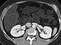
A computed tomography (CT) scan uses X-rays to make detailed pictures of structures inside of the body.
During the test, you will lie on a table that is attached to the CT scanner, which is a large doughnut-shaped machine. The CT scanner sends X-rays through the body area being studied. Each rotation of the scanner provides a picture of a thin slice of the organ or area. All of the pictures are saved as a group on a computer. They also can be printed.
In some cases, a dye called contrast material may be used. It may be put in a vein (IV) in your arm, or it may be placed into other parts of your body (such as the rectum or a joint) to see those areas better. For some types of CT scans, you drink the dye. The dye makes structures and organs easier to see on the CT pictures.
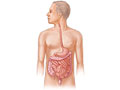
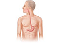
A CT scan can be used to study all parts of your body, such as the chest, belly, pelvis, or an arm or leg. It can take pictures of body organs, such as the liver, pancreas, intestines, kidneys, bladder, adrenal glands, lungs, and heart. It also can study blood vessels, bones, and the spinal cord.
Fluoroscopy CT is a special test that is not widely available. It uses a steady beam of X-rays to look at movement within the body. It allows the doctor to see your organs move or to guide a biopsy needle or other instrument into the right place inside your body.
Why It Is Done
CT scans are used to study areas of the body and the arms or legs.
- Chest (thorax). A CT scan of the chest can look for problems with the lungs, the heart, the esophagus, or the major blood vessel (aorta) or the tissues in the center of the chest. Some common chest problems a CT scan may find include infection, lung cancer, a pulmonary embolism, and an aneurysm. It also can be used to see if cancer has spread into the chest from another area of the body.
- Abdomen. A CT scan of the abdomen can find cysts, abscesses, infection, tumors, an aneurysm, enlarged lymph nodes, foreign objects, bleeding in the belly, diverticulitis, inflammatory bowel disease, and appendicitis.
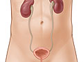
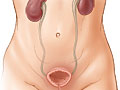
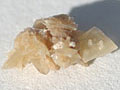
- Urinary tract. A CT scan of the kidneys, ureters, and bladder is called a CT KUB or CT urogram. This type of scan can find kidney stones, bladder stones, or blockage of the urinary tract. A special type of CT scan, called a CT intravenous pyelogram (IVP), uses injected dye (contrast material) to look for kidney stones, blockage, growths, infection, or other diseases of the urinary tract.
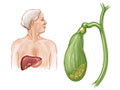
- Liver. A CT scan can find liver tumors, bleeding from the liver, and liver diseases. A CT scan of the liver can help determine the cause of jaundice.
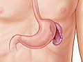
- Pancreas. A CT scan can find a tumor in the pancreas or inflammation of the pancreas (pancreatitis).
- Gallbladder and bile ducts. A CT scan can be used to check for blockage of the bile ducts. Gallstones occasionally show up on a CT scan. But other tests, such as ultrasound, usually are used to find problems with the gallbladder and bile ducts.
- Adrenal glands. A CT scan can find tumors or enlarged adrenal glands.
- Spleen. A CT scan can be used to check for an injury to the spleen or the size of the spleen.
- Pelvis. A CT scan can look for problems of organs in the pelvis. For a woman, these include the uterus, ovaries, and fallopian tubes. For a man, the pelvic organs include the prostate gland and the seminal vesicles.
- Arm or leg. A CT scan can look for problems of the arms or legs, including the shoulder, elbow, wrist, hand, hip, knee, ankle, or foot.
Other uses for a CT scan
A CT scan may be used to make sure a procedure is done correctly. For example, the doctor may use CT to guide a needle during a tissue biopsy or to guide the proper placement of a needle to drain an abscess.
For people with cancer, a CT scan can help determine how much the cancer has spread. This is called staging the cancer.
How To Prepare
Before the CT scan, tell your doctor if you:
- Are or might be pregnant.
- Are allergic to any medicines, including iodine dyes.
- Have a heart condition, such as heart failure.
- Have diabetes.
- Take metformin. You may have to adjust your medicine for a day before and after the test.
- Have had kidney problems.
- Have asthma.
- Have had multiple myeloma.
- Have had an X-ray test using barium contrast material (such as a barium enema) in the past 4 days. Barium shows up on X-ray films and makes it hard to see the picture clearly.
- Become very nervous in small spaces. You need to lie still inside the CT scanner, so you may need a medicine (sedative) to help you relax.
Arrange for someone to take you home in case you get a medicine to help you relax (sedative) for the test.
If you have a CT scan of your belly, you may be asked to not eat any solid foods starting the night before your scan. For a CT scan of the belly, you may drink contrast material. For some CT scans, you may need a laxative or an enema before the test.
Talk to your doctor about any concerns you have regarding the need for the test, its risks, how it will be done, or what the results will mean. To help you understand the importance of this test, fill out the medical test information form( What is a PDF document? ).
How It Is Done
A CT scan is usually done by a radiology technologist. The pictures are usually read by a radiologist, who writes the report. Other doctors also may review a CT scan.
You may need to take off any jewelry. You will need to take off all or most of your clothes, depending on which area is studied. You may be able to wear your underwear for some scans. You will be given a gown to use during the test.
During the test, you will lie on a table that is attached to the CT scanner.
The table slides into the round opening of the scanner, and the scanner moves around your body. The table will move while the scanner takes pictures. You may hear a click or buzz as the table and scanner move. It is very important to lie still during the test.
During the test, you may be alone in the scan room. But the technologist will watch you through a window. You will be able to talk to the technologist through a two-way intercom.
The test will take about 30 to 60 minutes. Most of this time is spent getting ready for the scan. The actual scan only takes a few seconds.
How It Feels
The test will not cause pain. The table you lie on may feel hard, and the room may be cool. It may be hard to lie still during the test.
Some people feel nervous inside the CT scanner.
If a medicine to help you relax (sedative) or dye (contrast material) is used, an IV is usually put in your hand or arm. You may feel a quick sting or pinch when the IV is started. The dye may make you feel warm and flushed and give you a metallic taste in your mouth. Some people feel sick to their stomachs or get a headache. Tell the technologist or your doctor how you are feeling.
Risks
The chance of a CT scan causing a problem is small.
- There is a chance of an allergic reaction to the dye (contrast material).
- If you breastfeed and are concerned about whether the dye used in this test is safe, talk to your doctor. Most experts believe that very little dye passes into breast milk and even less is passed on to the baby. But if you prefer, you can store some of your breast milk ahead of time and use it for a day or two after the test.
- If you have diabetes or take metformin (Glucophage), the dye may cause problems. Your doctor will tell you when to stop taking metformin and when to start taking it again after the test so you will not have problems.
- There is a small chance of getting cancer from some types of CT scans.footnote 1 The risk is higher in children, young adults, and people who have many radiation tests. If you are concerned about this risk, talk to your doctor about the benefits and risks of a CT scan, and confirm that the test is needed.
Results
A computed tomography (CT) scan uses X-rays to make detailed pictures of structures inside the body.
Complete results usually are ready for your doctor in 1 to 2 days.
|
Normal: |
The organs and blood vessels are normal in size, shape, and location. No blood vessels are blocked. |
|
No foreign objects (such as metal or glass fragments), growths (such as cancer), inflammation, or infection are present. |
|
|
No bleeding or collections of fluid are present. |
|
|
Abnormal: |
An organ is too large or too small, damaged, or infected. Abscesses are present. |
|
Foreign objects (such as metal or glass fragments) are present. |
|
|
Kidney stones or gallstones are present. |
|
|
Growths (such as tumors) are seen in the colon, lungs, ovaries, liver, bladder, kidneys, adrenal gland, or pancreas. |
|
|
A CT scan of the chest shows a pulmonary embolism, fluid in the lungs, or infection. |
|
|
An aneurysm is present. |
|
|
Blockage is found in the intestines or in the bile ducts. |
|
|
A CT of the belly shows inflammatory bowel disease or diverticulitis. |
|
|
Lymph nodes are enlarged. |
|
|
One or more blood vessels are blocked. |
|
|
A growth, fracture, infection, or other problem is found in an arm or leg. |
What Affects the Test
The following may stop you from having the test or may change the test results:
- Pregnancy. CT scans are not usually done during pregnancy.
- Barium used for another test. Barium shows up on a CT scan. If a CT scan of the belly is needed, it should be done before any tests that use barium, such as a barium enema.
- Metal objects in the body. These items, such as surgical clips or metal in joint replacements, may prevent a clear view of the body area.
- You are not able to lie still during the test.
What To Think About
- Sometimes your CT test results may be different than those from other types of X-ray tests, magnetic resonance imaging (MRI), or ultrasound scans because the CT scan provides a different view.
- An ultrasound test, which doesn’t use dangerous radiation, may give results similar to those of a CT scan. If you are concerned about radiation exposure, ask your doctor if you can have an ultrasound instead of a CT scan.
- Children who need a CT scan may need special instructions for the test. The child will likely need to hold his or her breath during the scan. If the child is too young to hold still or is afraid, the doctor may give the child a medicine (sedative) to help him or her relax.
- If your child is scheduled for a CT scan, talk with your child’s doctor about the need for the scan and the risk of radiation exposure to your child.
- Special CT scanners called spiral (helical) CT scanners and multi-slice (or multi-detector) CT scanners are sometimes used for this test. Many modern scanners are multi-slice scanners. These scanners can be used for many conditions, such as finding kidney stones, a pulmonary embolism, an enlarged prostate gland, or atherosclerosis. These special CT scanners can:
- Take better pictures of blood vessels and organs so other imaging tests may not be needed.
- Complete scans and provide pictures in less time.
- CT results are often compared to positron emission tomography (PET) results to help find cancer. Some new scanners do both scans at the same time.
- An electron beam CT scan is another type of CT scan that can find atherosclerosis and coronary artery disease. An electron beam CT scan is much faster than a standard CT scan and can take a good picture of a coronary artery while the heart is beating. Electron beam CT scans are not widely available. Another type of CT scanner, the multi-slice CT scan, is nearly as fast as electron beam CT scanners and is more widely available.
- A CT angiogram can show two- and three-dimensional pictures of blood vessels and the heart.
- Coronary calcium scans use a CT scan to check for early signs of coronary artery disease. This test is not advised for routine screening.
- MRI may give different information than a CT scan about certain conditions. To learn more, see the topic Magnetic Resonance Imaging (MRI).
- A low-dose CT is a way to scan the lungs more quickly than with a standard CT. Some doctors recommend this to screen for lung cancer in people who are older than 55 and who are at high risk for lung cancer.
- Experts disagree about the use of a CT method called full-body scanning to screen for coronary artery disease and cancers. Full-body scanning is expensive, can lead to unnecessary tests or surgery, and may increase the chance of cancer from the radiation exposure. Most doctors do not recommend these studies unless a person has a specific risk for a certain disease.
References
Citations
- Einstein AJ, et al. (2007). Estimating risk of cancer associated with radiation exposure from 64-slice computed tomography coronary angiography. JAMA, 298(3): 317–323.
Other Works Consulted
- Bluemke, D, et al. (2008). Noninvasive coronary artery imaging: Magnetic resonance angiography and multidetector computed tomography angiography. A scientific statement From the American Heart Association Committee on Cardiovascular Imaging and Intervention of the Council on Cardiovascular Radiology and Intervention, and the Councils on Clinical Cardiology and Cardiovascular Disease in the Young. Circulation, 118: 586–606.
- Detterbeck FC, et al. (2013). Screening for lung cancer. Diagnosis and management of lung cancer, 3rd ed. American College of Chest Physicians evidence-based clinical practice guidelines. Chest, 143(5, Suppl): e78S–e92S.
- Fischbach FT, Dunning MB III, eds. (2009). Manual of Laboratory and Diagnostic Tests, 8th ed. Philadelphia: Lippincott Williams and Wilkins.
- National Comprehensive Cancer Network (2010). Non–Small Cell Lung Cancer, version 2.2010. Available online: http://www.nccn.org/professionals/physician_gls/f_guidelines.asp.
- Pagana KD, Pagana TJ (2010). Mosby’s Manual of Diagnostic and Laboratory Tests, 4th ed. St. Louis: Mosby.
- Pearce MS, et al. (2012). Radiation exposure from CT scans in childhood and subsequent risk of leukaemia and brain tumours: A retrospective cohort study. Lancet, 380(9840): 499–505.
- U.S. Food and Drug Administration (2008). FDA preliminary public health notification: Possible malfunction of electronic medical devices caused by computed tomography (CT) scanning. Available online: http://www.fda.gov/MedicalDevices/Safety/AlertsandNotices/PublicHealthNotifications/ucm061994.htm.
Current as of: March 28, 2019
Author: Healthwise Staff
Medical Review:Adam Husney MD – Family Medicine & E. Gregory Thompson MD – Internal Medicine & Martin J. Gabica MD – Family Medicine & Howard Schaff MD – Diagnostic Radiology

This information does not replace the advice of a doctor. Healthwise, Incorporated, disclaims any warranty or liability for your use of this information. Your use of this information means that you agree to the <a class=”HwLinkExternal” href=”https://www.healthwise.org/spec