Doppler Ultrasound
Test Overview
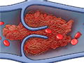
A Doppler ultrasound test uses reflected sound waves to see how blood flows through a blood vessel. It helps doctors assess the blood flow through major arteries and veins, such as those of the arms, legs, and neck. It can show blocked or reduced flow of blood through narrow areas in the major arteries of the neck. This problem could cause a stroke. The test also can find blood clots in leg veins (deep vein thrombosis, or DVT) that could break loose and block blood flow to the lungs. This problem is called a pulmonary embolism. During pregnancy, Doppler ultrasound may be used to look at blood flow in an unborn baby to check the baby’s health.
During Doppler ultrasound, a handheld device is passed lightly over the skin above a blood vessel. The device is called a transducer. It sends and receives sound waves that are amplified through a microphone. The sound waves bounce off solid objects, including blood cells. The movement of blood cells causes a change in the pitch of the reflected sound waves. This is called the Doppler effect. If there is no blood flow, the pitch does not change.
Information from the reflected sound waves can be used to make graphs or pictures that show the flow of blood through the blood vessels. These graphs or pictures can be saved and reviewed later.
The three basic types of Doppler ultrasound are:
- “Bedside” or continuous wave Doppler. This type uses the change in pitch of the sound waves to provide information about blood flow through a blood vessel. The doctor listens to the sounds made by the transducer to assess the blood flow through an area that may be blocked or narrowed. This type of ultrasound can be done at the bedside in the hospital. It uses a portable machine that can quickly check the extent of blood vessel damage or disease.
- Duplex Doppler. This test uses standard ultrasound methods to make a picture of a blood vessel and the organs around it. A computer turns the Doppler sounds into a graph. This graph helps to show the speed and direction of blood flow through the blood vessel.
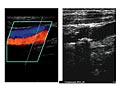
- Color Doppler. Color Doppler uses standard ultrasound methods to make a picture of a blood vessel. A computer changes the Doppler sounds into colors that are overlaid on the image of the blood vessel. These colors show the speed and direction of blood flow through the vessel. Power Doppler is a special type of color Doppler. Power Doppler can get some images that are hard or impossible to get using standard color Doppler. Power Doppler is most often used to look at blood flow through vessels within solid organs.
Why It Is Done
Doppler ultrasound is done to:
- Find blood clots and blocked or narrowed blood vessels in almost any part of the body. It’s most often used for the neck, arms, and legs.
- Check leg pain that may be caused by intermittent claudication. This is a condition caused by atherosclerosis of the lower limbs.
- Assess blood flow after a stroke or other condition that might be caused by a problem with blood flow. After a stroke, this can be done with a test called transcranial Doppler (TCD) ultrasound.
- Check for varicose veins or other vein problems.
- Map veins that may be used for blood vessel grafts. It also can look at the health of grafts used to bypass blockage in an arm or leg.
- Find out the amount of blood flow to a transplanted kidney or liver.
- Monitor the flow of blood after blood vessel surgery.
- Find the presence, amount, and location of arterial plaque. Plaque in the carotid arteries can reduce blood flow to the brain. This may increase the risk of stroke.
- Guide treatment such as laser or radiofrequency ablation of abnormal veins.
- Check the health of a fetus. It may check blood flow in the umbilical cord, through the placenta, or in the heart and brain of the fetus. This test can show if the fetus is getting enough oxygen and nutrients. Doppler ultrasound may be used to guide decisions during pregnancy when:
- The fetus is smaller than normal for the gestational age. Blood flow through the large blood vessel in the umbilical cord (the umbilical artery) can be looked at.
- Rh sensitization has occurred. Blood flow through a blood vessel in the brain can be used to check the health of the fetus.
- The mother has other problems, such as preeclampsia or sickle cell disease.
A transcranial Doppler (TCD) ultrasound may be used in children with sickle cell disease. The test can check for the risk of stroke.
How To Prepare
For 30 minutes to 2 hours before the test, you may need to stop using products that contain nicotine. This includes cigarettes and chewing tobacco. Nicotine causes blood vessels to narrow, which may give false results.
How It Is Done
This test is done by a doctor who is an expert in imaging tests (radiologist). Or it may be done by an ultrasound technologist (sonographer) who works with a radiologist. It is done in an ultrasound room in a hospital or doctor’s office.
You will need to remove any jewelry that might affect the Doppler ultrasound scan. You may need to take off all or most of your clothes, depending on which part of the body is being examined. You may be allowed to keep on your underwear if it does not affect the test. You will be given a cloth or paper covering to use during the test.
- For abdominal scans, you will lie on your back.
- For chest scans, you will lie on your back with your neck slightly extended.
- For head and neck scans, your head may be turned to one side.
- For an arm or leg scan, your head will be slightly raised. The exposed arm or leg will be turned slightly outward. Sometimes for a leg scan, you may be asked to lie on your stomach.
- During pregnancy, you will lie on your back or on your left side with your belly exposed.
Gel is applied to the skin to help the sound waves pass through. The transducer is placed in the gel and moved along the skin. You need to lie very still during the test. You may hear sounds from the flow of blood through the blood vessels.
The test usually takes 30 to 60 minutes.
Arteries in the arms and legs
This test is often done on both arms or both legs. Even if the suspected blood flow problem is in only one limb, both may be tested to compare them. If your arms are being tested, they will be tested first while you lie down. Then they’ll be tested again while you sit.
Depending on which blood vessels are being tested, a blood pressure cuff may be wrapped around one or both limbs. It allows your blood pressure to be taken at several different places. When the legs are tested, a blood pressure cuff may be wrapped first around the calf and then around the thigh. The test may be done at several places on your leg. When the arms are tested, the pressure cuff may be wrapped first around the forearm and then around the upper arm.
Testing may be done before and after exercise, if you are healthy enough.
Veins in the arms and legs
For this test, you will be asked to lie down and breathe normally. You must lie very still. Any changes in blood flow that are affected by how you breathe are noted.
The test may be repeated while the examiner presses on the veins close to the surface of your skin. This helps to find a clot in the vein. It’s called a compression maneuver. The examiner may do this maneuver with your legs or arms in different positions. This is to make sure that the blood supply is not blocked in these positions. He or she may also squeeze your calf or forearm to help blood move more quickly through the veins. This is called an augmentation maneuver. It is done to check blood flow toward your heart.
While your legs are being tested, you may also be asked to try to breathe out strongly with your nose pinched and your mouth closed. This is called a Valsalva maneuver. It usually causes a sudden change in blood flow through the veins.
Arteries in the neck
You will be asked to lie down with a pillow under your head for support. The test is done on both sides of your neck. Then the results are compared to standard values to find out how much the arteries are blocked or narrowed.
Transcranial ultrasound
For this type of ultrasound, the transducer is passed lightly over the skin at the base or side of your skull.
During pregnancy
The transducer is moved back and forth on your belly until the doctor finds the blood vessel that needs to be studied. After the doctor has found the blood vessel, it may take some time to assess the blood flow.
How It Feels
Having a Doppler ultrasound test doesn’t normally cause discomfort. The gel may feel cold when it is put on your skin unless it is warmed first. If your blood pressure is taken during the test, you will feel pressure when the blood pressure cuff is inflated.
Risks
There are no known risks linked with a Doppler ultrasound test. This test will not harm an unborn baby (fetus).
Results
A Doppler ultrasound test uses reflected sound waves to see how blood flows through a blood vessel.
|
Normal: |
The test does not show significant narrowing or other problems in any of the arteries. |
|
There is no sign of a clot in any of the veins examined. The size and position of veins are normal. |
|
|
Normal blood flow is found in the blood vessels that supply oxygen and nutrients to a fetus. |
|
|
Abnormal: |
For continuous wave Doppler or duplex Doppler, differences in blood flow between the right and left sides of the body may be heard. At the exact spot where an artery is blocked or narrowed, the sound may be high-pitched or turbulent. Blockage (such as from a blood clot), an aneurysm, or narrowing of a blood vessel may be found. The speed of blood flow may be compared to standard values to find out how blocked or narrow the blood vessel is. |
|
A duplex Doppler ultrasound graph may show blood flow that isn’t normal. This is a sign of a blocked or narrowed blood vessel. |
|
|
A color Doppler image may show a blocked or narrowed blood vessel or an aneurysm. |
|
|
In the veins, a blood clot may be present if blood flow does not change in response to breathing or does not increase after either a compression maneuver or Valsalva maneuver. Incomplete blockage of a vein by a blood clot may be seen on color Doppler or during a compression maneuver. |
|
|
Abnormal veins, such as varicose veins, are seen. |
|
|
There is an abnormal increase or decrease in blood flow through the vessels that supply oxygen and nutrients to a fetus. |
What Affects the Test
You may not be able to have the test, or the results may not be helpful, if:
- There are bones above the area being studied or gas in the intestines. This may affect the pictures.
- You are not able to stay still during the test.
- You are extremely overweight.
- You have abnormal heart rhythms (arrhythmias) or heart disease. This may cause changes in blood flow patterns even though the blood vessels are normal.
- Your arm or leg is cold. Blood flow through that limb may be slowed.
- You have an open wound in the area that needs to be viewed.
What To Think About
- It takes a skilled examiner to get accurate test results for a Doppler ultrasound. The scans are usually read within a short period of time in case repeat tests are needed.
- Doppler ultrasound requires a person to hold very still, so some children may need to be given medicine to make them sleep. This is done so that their movements do not affect the results.
- Angiography and venography are X-ray tests that use an injected contrast material. In many cases, Doppler ultrasound may be done instead of these tests. This is because it’s faster, costs less, and is noninvasive. If results from a Doppler ultrasound are unclear, an angiography or venography test may be done. Angiography is usually more accurate than Doppler ultrasound. It is also considered the most reliable test for checking the blood flow through an artery. Magnetic resonance angiography (MRA) and computed tomography angiography (CTA) may be done instead of regular angiography. That’s because these tests are less invasive and easier to do than regular angiography. In some cases, venography may be needed to confirm a suspected vein problem.
- A Doppler ultrasound may be used to check many body organs. It’s also safe to have it when you are pregnant.
References
Other Works Consulted
- Chernecky CC, Berger BJ (2008). Laboratory Tests and Diagnostic Procedures, 5th ed. St. Louis: Saunders.
- Fischbach FT, Dunning MB III, eds. (2009). Manual of Laboratory and Diagnostic Tests, 8th ed. Philadelphia: Lippincott Williams and Wilkins.
- Pagana KD, Pagana TJ (2010). Mosby’s Manual of Diagnostic and Laboratory Tests, 4th ed. St. Louis: Mosby Elsevier.
- Roman AS (2013). Late pregnancy complications. In AH DeCherney et al., eds., Current Diagnosis and Treatment Obstetrics & Gynecology, 11th ed., pp. 250–266. New York: McGraw-Hill.
Current as of: March 28, 2019
Author: Healthwise Staff
Medical Review:Kathleen Romito, MD – Family Medicine & Adam Husney, MD – Family Medicine & Martin J. Gabica, MD – Family Medicine & Howard B. Schaff, MD – Diagnostic Radiology

This information does not replace the advice of a doctor. Healthwise, Incorporated, disclaims any warranty or liability for your use of this information. Your use of this information means that you agree to the Terms of Use. Learn how we develop our content.