Magnetic Resonance Angiogram (MRA)
Test Overview
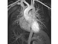
A magnetic resonance angiogram (MRA) is a type of magnetic resonance imaging (MRI) scan that uses a magnetic field and pulses of radio wave energy to provide pictures of blood vessels inside the body. In many cases MRA can provide information that can’t be obtained from an X-ray, ultrasound, or computed tomography (CT) scan.
MRA can find problems with the blood vessels that may be causing reduced blood flow. With MRA, both the blood flow and the condition of the blood vessel walls can be seen. The test is often used to look at the blood vessels that go to the brain, kidneys, and legs. Information from an MRA can be saved and stored on a computer for further study. Photographs of selected views can also be made.
During MRA, the area of the body being studied is placed inside an MRI machine. Contrast material is often used during MRA to make blood vessels show up more clearly.
Why It Is Done
A magnetic resonance angiogram (MRA) is done to look for:
- A bulge (aneurysm), clot, or the buildup of fat and calcium deposits (stenosis caused by plaque) in the blood vessels leading to the brain.
- An aneurysm or tear (dissection) in the aorta, which carries blood from the heart to the rest of the body.
- Narrowing (stenosis) of the blood vessels leading to the heart, lungs, kidneys, or legs.
How To Prepare
Before a magnetic resonance angiogram (MRA), tell your doctor and the MRI technologist if you:
- Are allergic to any medicines. The contrast material used for MRA does not contain iodine. If you know that you are allergic to the contrast material used for MRA, tell your doctor before having another test.
- Are or might be pregnant.
- Have any metal implanted in your body. This information helps your doctor know if the test is safe for you. Tell your doctor if you have:
- Heart and blood vessel devices such as a coronary artery stent, pacemaker, ICD (implantable cardioverter-defibrillator), or metal heart valve.
- Metal pins, clips, or metal parts in your body, including artificial limbs and dental work or braces.
- Any other implanted medical device, such as a medicine infusion pump.
- Cosmetic metal implants, such as in your ears.
- Have an intrauterine device (IUD) in place. An IUD may prevent you from having the MRA test done.
- Become very nervous in small spaces. You need to lie very still inside the MRI machine, so you may need to have the test done with open MRI equipment. It is not as confining as standard MRI machines. You may need medicine to help you relax. Some blood vessels may not be seen clearly with an open MRI scanner.
- Have any other health conditions, such as kidney problems or sickle cell anemia, that may prevent you from having an MRA using contrast material.
- Wear any medicine patches. The MRI may cause a burn at the patch site.
For some abdominal MRI tests, you may be asked to not eat or drink before the test.
You may need to arrange for someone to drive you home after the test, if you are given a medicine (sedative) to help you relax.
If you are having blood flow studies, do not use tobacco products and do not eat or drink (including alcohol or caffeinated beverages) for 2 hours before the test. Do not take iron supplements on the day of the test.
You may be asked to sign a consent form that says you understand the risks of the test and agree to have it done.
Talk to your doctor about any concerns you have regarding the need for the test, its risks, how it will be done, or what the results will mean. To help you understand the importance of this test, fill out the medical test information form( What is a PDF document? ).
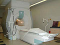
How It Is Done
A magnetic resonance angiogram (MRA) test is usually done by a magnetic resonance image (MRI) technologist. The test is done in an MRI machine. The resulting pictures are usually interpreted by a radiologist. But some other types of doctors can also interpret an MRA scan.
Before the test
- Remove all metal objects, such as hearing aids, dentures, jewelry, watches, hairpins, wigs, and eyeglasses, from your body because these objects may be attracted to the powerful magnet used for the test. If you have had a car crash or an accident while working with metal, it is possible that you have metal fragments in your head, eyes, skin, or spine. An X-ray may be taken before the MRA to see if you can have the test.
- Take off all or most of your clothes, depending on which area is examined. You may be allowed to keep on your underwear if it does not get in the way. You will be given a cloth or paper covering to use during the test.
- You may be given a sedative if you are nervous or you do not think you can lie still for the test.
During the test
- You will lie on your back on a table that is part of the MRI scanner.
- If you are cold or uncomfortable, you may want to ask for a pillow or blanket.
- Your head, chest, and arms may be held with straps to help you remain still.
- The table will slide into a space that contains the magnet. Depending on the part of your body to be examined, your head, limbs (such as your legs), or your entire body will be moved into the center of the magnet.
- Inside the scanner, you may hear a fan and feel air moving. You may also hear tapping or thumping noises as the MRA scans are taken. You may want to ask for ear plugs to reduce the noise.
- It is important to hold completely still while the scan is being done. Otherwise, repeat scans may be needed.
- You may be asked to hold your breath for short periods of time.
- You may be alone in the scanner room. But the technologist will watch you through an observation window, and you will be able to talk to him or her through an intercom.
If contrast material is needed, the technologist will put it in an IV in your arm. The material may be given over 1 to 2 minutes. Then more MRI scans are done.
An MRA test usually takes 30 to 90 minutes but can take as long as 2 hours.
How It Feels
You won’t have pain from the magnetic field or radio waves used for the MRI test. The table you lie on may feel hard and the room may be cool. You may be tired or sore from lying in one position for a long time.
Some people feel discomfort or anxiety (claustrophobia) inside the MRI magnet. If this keeps you from lying still, you can be given a sedative to help you relax. Open MRI machines are less confining than standard MRI and may be helpful if you are claustrophobic.
If a contrast material is used, you may feel some coolness when it is put into your IV. In rare cases, you may feel:
- A tingling feeling in the mouth if you have metal dental fillings.
- Warmth in the area being examined. This is normal. Tell the technologist if you have nausea, vomiting, headache, dizziness, pain, burning, or breathing problems.
Risks
There is a slight risk of having an allergic reaction if contrast material is used during the MRA scan. Most reactions can be controlled using medicine.
An MRI can cause a burn with some medicine patches. Be sure to tell your doctor if you are wearing a patch.
A dye (contrast material) that contains gadolinium may be used in this test. Be sure to tell your doctor if:
- You are pregnant or think you may be pregnant.
- You have kidney problems.
- You’ve had more than one test that used gadolinium.
The U.S. Food and Drug Administration (FDA) has safety warnings about gadolinium. But for most people, the benefit of its use in this test outweighs the risk.
If you breastfeed and are concerned about whether the dye used in this test is safe, talk to your doctor. Most experts believe that very little dye passes into breast milk and even less is passed on to the baby. But if you prefer, you can store some of your breast milk ahead of time and use it for a day or two after the test.
Results
A magnetic resonance angiogram (MRA) is a type of magnetic resonance imaging (MRI) scan that uses a magnetic field and pulses of radio wave energy to provide pictures of blood vessels inside the body. The radiologist may talk to you about the results of your MRA right after the test. Complete results are usually available for your doctor in 1 to 2 days.
|
Normal: |
The blood vessels look normal and the blood flow through them is not reduced or stopped. No blood clots or large plaque buildup is seen. |
|
Blood vessel walls are normal. No bleeding, abnormal collections of fluid, blockage in the flow of blood, or bulges in the blood vessels (aneurysms) are present. |
|
|
Abnormal: |
Partial or complete blockage of a blood vessel may be seen. Blockage may be caused by a blood clot, the buildup of fat and calcium deposits (plaque), or narrowing (stenosis) of the blood vessel. |
|
A bulge (aneurysm) in the blood vessel wall may be seen. Damage to the wall of a blood vessel may be seen. |
Conventional angiogram or a CT angiogram (computed tomography angiogram) may be needed after MRA if a problem, such as an aneurysm, is found or if surgery may be needed.
What Affects the Test
Reasons you may not be able to have the test or why the results may not be helpful include:
- Pregnancy. Although the strong magnetic field used for an MRA does not appear to be harmful, MRA usually is not done when you are pregnant. If a view of your belly is needed and you are pregnant, an ultrasound test may be done instead.
- You are using a medical device that contains metal, such as an IUD, a pacemaker, some types of artificial limbs, or medicine infusion pumps. These devices can malfunction or cause problems during an MRA scan.
- Not being able to lie still during the test. The results of MRA may not be accurate if you can’t remain still during the test.
- Being overweight and not fitting into the opening of some MRI scanners.
What To Think About
- While MRA is a safe and valuable test for looking at blood vessels inside the body, it is more expensive than other imaging techniques, and it may not be available in all medical centers.
- An advantage of MRA is that no radiation is involved.
- You may be able to have your MRA in an open MRI machine that doesn’t enclose your entire body. But open MRI machines aren’t available everywhere. The pictures from an open MRI may not be as good as those from a standard MRI machine. Also, these machines may not be able to do all the studies needed to check for problems.
- Open MRI is useful for people who are claustrophobic or obese.
- Conventional angiogram or a CT angiogram (computed tomography angiogram) may be done to double-check abnormal results from the MRA in some types of blood vessels such as the aorta. These tests might be an option especially if surgery is being considered to treat the problem.
- MRA results may show an aneurysm is present when it is not (false-positive). It also may show no aneurysm when one is present (false-negative). MRA is most accurate for larger blood vessels.
Current as of: September 26, 2018
Author: Healthwise Staff
Medical Review:Rakesh K. Pai, MD – Cardiology, Electrophysiology & E. Gregory Thompson, MD – Internal Medicine & Adam Husney, MD – Family Medicine & Kathleen Romito, MD – Family Medicine & George J. Philippides, MD, FACC – Cardiology

This information does not replace the advice of a doctor. Healthwise, Incorporated, disclaims any warranty or liability for your use of this information. Your use of this information means that you agree to the Terms of Use. Learn how we develop our content.