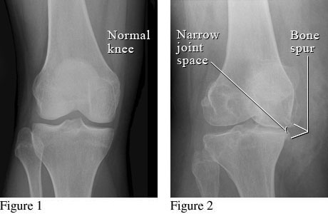
X-Ray of Osteoarthritis of the Knee
Current as of: April 1, 2019
Author: Healthwise Staff
Medical Review:Anne C. Poinier, MD – Internal Medicine & Martin J. Gabica, MD – Family Medicine & Kathleen Romito, MD – Family Medicine & Stanford M. Shoor, MD – Rheumatology