Echocardiogram
Test Overview
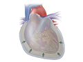
An echocardiogram (also called an echo) is a type of ultrasound test that uses high-pitched sound waves that are sent through a device called a transducer. The device picks up echoes of the sound waves as they bounce off the different parts of your heart. These echoes are turned into moving pictures of your heart that can be seen on a video screen.
The different types of echocardiograms are:
- Transthoracic echocardiogram (TTE). This is the most common type. Views of the heart are obtained by moving the transducer to different locations on your chest or abdominal wall.
- Stress echocardiogram. During this test, an echocardiogram is done both before and after your heart is stressed either by having you exercise or by injecting a medicine that makes your heart beat harder and faster. A stress echocardiogram is usually done to find out if you might have decreased blood flow to your heart (coronary artery disease).
- Doppler echocardiogram. This test is used to look at how blood flows through the heart chambers, heart valves, and blood vessels. The movement of the blood reflects sound waves to a transducer. The ultrasound computer then measures the direction and speed of the blood flowing through your heart and blood vessels. Doppler measurements may be displayed in black and white or in color.
- Transesophageal echocardiogram (TEE). For this test, the probe is passed down the esophagus instead of being moved over the outside of the chest wall. TEE shows clearer pictures of your heart, because the probe is located closer to the heart and because the lungs and bones of the chest wall do not block the sound waves produced by the probe. A sedative and an anesthetic applied to the throat are used to make you comfortable during this test.
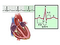
Echo can be used as part of a stress test and with an electrocardiogram (EKG or ECG) to help your doctor learn more about your heart.
Why It Is Done
Transthoracic echocardiogram (TTE)
This test is done to:
- Look for the cause of abnormal heart sounds (murmurs or clicks), an enlarged heart, unexplained chest pain or pressure, shortness of breath, or irregular heartbeats.
- Check the thickness and movement of the heart wall.
- Look at the heart valves and check how well they work.
- See how well an artificial heart valve is working.
- Measure the size and shape of the heart’s chambers.
- Check the ability of your heart chambers to pump blood (cardiac performance). During an echocardiogram, your doctor can calculate how much blood your heart is pumping during each heartbeat (ejection fraction). You might have a low ejection fraction if you have heart failure.
- Detect a disease that affects the heart muscle and the way it pumps, such as cardiomyopathy.
- Look for blood clots and tumors inside the heart.
A transthoracic echocardiogram may also be used to:
- Look for congenital heart defects or to check the effectiveness of previous surgery to repair a congenital heart defect.
- Check how well your heart works after a heart attack.
- Identify the specific cause of heart failure.
- Look for a collection of fluid around the heart (pericardial effusion).
- Look for a thickening of the lining (pericardium) around the heart.
Stress echocardiogram
A stress echo may be done to:
- Identify and monitor reduced blood flow to heart muscle (ischemia). This is usually more apparent after some form of stress, such as exercise or medicine.
Doppler echocardiogram
A Doppler echocardiogram can be done during a transthoracic echocardiogram (TTE), a transesophageal echocardiogram (TEE), or a stress echocardiogram to:
- Measure the speed at which blood travels through the heart.
- Measure the blood pressure and speed of blood flow through the heart valves.
Transesophageal echocardiogram (TEE)
Transesophageal echocardiogram (TEE) may be done to:
- Monitor heart function during surgery.
- Check how well an artificial heart valve works.
- Look for masses or blood clots in the upper left chamber (left atrium) of the heart.
- Identify abnormal blood flow between the chambers of the heart (cardiac shunt).
- Help find out if you have endocarditis, which is an infection of the heart’s valves or its inner lining (endocardium).
- Guide procedures done during cardiac catheterization.
- Help find out if you have a tear in the aorta (aortic dissection).
How To Prepare
Transthoracic echocardiogram (TTE) and Doppler echocardiogram
You do not need any special preparation for a transthoracic or Doppler echocardiogram.
Stress echocardiogram
Do not eat heavily for a few hours before a stress echo to help prevent nausea. You may feel nauseated if you exercise with a full stomach or from the injection of dobutamine.
Wear flat, comfortable shoes (no bedroom slippers or sandals) and loose, lightweight shorts or sweatpants for an exercise stress echo.
Ask your doctor whether you should take your regular medicines as usual. Tell your doctor if you take insulin.
Transesophageal echocardiogram (TEE)
Do not eat or drink for at least 6 hours before the TEE.
If you have dentures or dental prostheses, you may need to remove them before the test.
If you have medical problems involving the throat, esophagus, or stomach, tell your doctor before getting this test.
Before a TEE, you will be given a sedative. You will not be able to drive for at least 12 hours after the procedure. Be sure to make arrangements in advance for someone to pick you up after the test.
Before an echocardiogram, you may be asked to sign a consent form that says you understand the risks of the test and agree to have it done.
Talk to your doctor about any concerns you have regarding the need for the test, its risks, how it will be done, or what the results will mean. To help you understand the importance of this test, fill out the medical test information form( What is a PDF document? ).
How It Is Done
An echocardiogram may be done in a hospital, clinic, or doctor’s office. It can also be done at your bedside in the hospital.
You will need to remove any jewelry and clothes above your waist (you may be allowed to keep on your underwear if it does not interfere with the test). You may be given a cloth or paper covering to use during the test.
A transthoracic echocardiogram (TTE), Doppler echocardiogram, and stress echocardiogram are performed by a specially trained ultrasound technician. A transesophageal echocardiogram (TEE) is performed by a cardiologist with the help of assistants.
You may receive an IV so you can get medicine during the test. The IV can be used to give you a contrast material, which helps your doctor check your heart function. A contrast material may be used if it is difficult to get good views of your heart. A good view might be hard to get because of certain conditions such as obesity or chronic lung disease.
Transthoracic echocardiogram (TTE) and Doppler echocardiogram
You will lie on your back or on your left side on a bed or table. Small pads or patches (electrodes) will be taped to your arms and legs to record your heart rate during the test. To learn more, see Electrocardiogram.
A small amount of gel will be rubbed on the left side of your chest to help pick up the sound waves. A small instrument (transducer) that looks like a microphone is pressed firmly against your chest and moved slowly back and forth. This instrument sends sound waves into the chest and picks up the echoes as they reflect off different parts of the heart. The echoes are sent to a video monitor that records pictures of your heart for later viewing and evaluation. The room is usually darkened to help the technician see the pictures on the monitor.
At times you will be asked to hold very still, breathe in and out very slowly, hold your breath, or lie on your left side. The transducer is usually moved to different areas on your chest that provide specific views of your heart.
The test usually takes from 30 to 60 minutes. When the test is over, the gel is wiped off and the electrodes are removed.
Exercise stress echocardiogram
An echo without activity or stress will be done before you start exercising. This is called the baseline, and after it is established you will exercise for a specific amount of time. You will either walk on a treadmill or pedal a stationary bicycle while being monitored by an EKG machine. To learn more, see Exercise Electrocardiogram.
You will then lie on a bed or table, and another echocardiogram will be done. At times you will be asked to hold very still, breathe in and out very slowly, hold your breath, or lie on your left side. The transducer is usually moved to different areas on your chest that provide specific views of your heart.
An exercise stress echo takes about 30 to 60 minutes.
Dobutamine stress echocardiogram
Sometimes medicine called dobutamine is used instead of exercise to stress your heart. For this test, you will lie on your back or left side on a bed or exam table, and a baseline echocardiogram will be done. EKG electrodes will be taped to your arms and legs to record your heart rate during the test.
Next, the technician cleans the site on your arm where the medicine will be injected, and an intravenous (IV) line will be placed in a vein in your arm.
After the IV is started, you will be given dobutamine, which increases your heart rate and causes your heart to work harder. Echocardiogram images will be taken while you receive the dobutamine. Your peak heart rate is reached in about 15 minutes. At times you will be asked to hold very still, breathe in and out very slowly, hold your breath, or lie on your left side. After your peak heart rate is reached, the medicine will be stopped and your heart rate will return to normal (in about 1 to 3 minutes). More echocardiogram images will be taken when your heart rate returns to normal.
A dobutamine stress echo takes about an hour.
Transesophageal echocardiogram (TEE)
Before a TEE, your throat may be numbed with an anesthetic spray, gargle, or lozenge to relax your gag reflex and to ease insertion of the probe. Shortly before the procedure begins, an IV line will be placed in a vein in your arm. Medicine to decrease saliva and stomach secretions may be given through the IV. A pain medicine and sedative will be given to you through the IV in your arm during the procedure. You should feel relaxed and drowsy but still alert enough to cooperate.
Your heart rate, breathing rate, and blood pressure will be monitored throughout the procedure. Also, a small device used to measure the amount of oxygen in your blood (pulse oximeter) may be attached to your finger or earlobe.
You will be asked to lie on your left side with your head tilted slightly forward. A mouth guard may be inserted to protect your teeth from the probe. Then the lubricated tip of the probe will be guided into your mouth while your doctor gently presses your tongue out of the way. You may be asked to swallow to help move the tube along. It may be helpful to remember that the instrument is no thicker than many foods you swallow. When the probe is in your esophagus, it will be moved down gently to the level of your upper right heart chamber (atrium), and ultrasound images will be taken. You will not feel or hear the sound waves during the test.
During the procedure, try not to swallow unless requested. An assistant may remove the saliva from your mouth with a suction device, or you can just let the saliva drain from the side of your mouth. A transesophageal echo is generally painless, though you may feel nauseated and uncomfortable while the probe is in your throat.
The test takes about 2 hours. The probe will be in place in your esophagus for about 10 to 20 minutes.
How It Feels
Transthoracic echocardiogram (TTE) and Doppler echocardiogram
You will not have pain from the echocardiogram. Gel is put on your chest for the ultrasound. It may feel cool. The handheld ultrasound device is pressed firmly against your chest, but it does not cause pain. You will not hear or feel the sound waves.
You may feel uncomfortable from lying still or from the transducer pressing on your chest. If you need to take a break, tell the technician.
Most people do not experience any discomfort from ultrasound tests. But if you have severe difficulty breathing or cannot lie flat for a long exam, you may not be able to have an entire echo study. Talk to your doctor or the technician performing your echo about any concerns you have.
Dobutamine stress echocardiogram
- You may have a brief, sharp pain when the intravenous (IV) needle is placed in a vein in your arm.
- If medicine to stress your heart is used, you may have symptoms of mild nausea, headache, dizziness, flushing, or angina (such as chest pain or pressure). These symptoms only last a few minutes.
Transesophageal echocardiogram (TEE)
During the test
- You may notice a brief, sharp pain when the intravenous (IV) needle is placed in a vein in your arm.
- The anesthetic sprayed into your throat may taste bitter and will make your tongue and throat feel numb and swollen. Some people report that they feel as if they cannot breathe at times because of the probe in their throat, but this is a false sensation caused by the anesthetic. There is always plenty of breathing space around the probe in your mouth and throat. Remember to relax and take slow, deep breaths.
- You may gag and feel nauseous, bloated, or have mild belly cramps when the probe is moved. If the discomfort is severe, alert your doctor with an agreed-upon signal or a tap on the arm. Even though you won’t be able to talk during the procedure, you can still communicate.
- The IV medicines will make you feel sleepy. Other side effects—such as heavy eyelids, trouble speaking, a dry mouth, or blurred vision—may last for several hours after the test. You probably will not be able to remember much of the test.
After the test
- You may have a tickling, dry throat; slight hoarseness; or a mild sore throat. These symptoms may last for 2 to 3 days. Throat lozenges and warm saltwater gargles can help relieve these symptoms. Throat lozenges can be used by people age 4 or older. And most people can gargle at age 8 and older.
- Do not drink alcohol for 24 hours.
- Contact your doctor immediately if you have:
- Difficulty swallowing or talking.
- Shortness of breath or a fast heartbeat.
- Chest pain.
Risks
An echocardiogram is safe, because the test uses only sound waves to evaluate your heart. These high-frequency sound waves have not been shown to have any harmful effects.
If contrast material is used, there is a slight risk of having an allergic reaction. Most reactions can be controlled using medicine.
Transthoracic echocardiogram (TTE) and Doppler echocardiogram
There are no known risks from a transthoracic or Doppler echocardiogram. During a transthoracic echo, the technician may have to press hard on your chest with the transducer. Tell the technician if you feel any pain or discomfort.
Stress echocardiogram
A stress echocardiogram can cause dizziness, low blood pressure, shortness of breath, nausea, irregular heartbeats, and heart attack.
Transesophageal echocardiogram (TEE)
A transesophageal echocardiogram (TEE) can sometimes cause:
- Nausea.
- Mouth and throat discomfort.
- Minor bleeding.
- Trouble breathing.
- Slow or abnormal heartbeats.
Insertion of the probe may tear or puncture your esophagus. This is rare.
This test is not recommended if you have:
- Recently had radiation treatment to your neck or chest.
- Serious problems with your esophagus, such as a very narrow esophagus, dilated (engorged) veins in the esophagus that could rupture and bleed (esophageal varices), or severe arthritis of your neck.
- Trouble swallowing.
- A bleeding disorder, such as hemophilia.
Results
An echocardiogram is a type of ultrasound test that uses high-pitched sound waves that are sent through a device called a transducer. The device picks up echoes of the sound waves as they bounce off the different parts of your heart. These echoes are turned into moving pictures of your heart that can be seen on a video screen.
Results are usually available within one day. If the test is done by a cardiologist, the results may be available immediately after the test.
|
Normal: |
The heart chambers and walls of the heart are of normal size and thickness, and they move normally. |
|
Heart valves are working normally, with no leaks or narrowing. There is no sign of infection. |
|
|
The amount of blood pumped from the left ventricle with each heartbeat (ejection fraction) is normal. |
|
|
There is no excess fluid in the sac surrounding the heart, and the lining around the heart is not thickened. |
|
|
There are no tumors and blood clots in the heart chambers. |
|
|
Abnormal: |
Heart chambers are too big. The walls of the heart are thicker or thinner than normal. A thin heart wall may mean poor blood flow to the heart muscle or an old heart attack. A thin, bulging area of the heart wall may indicate a bulge in the ventricle (ventricular aneurysm). The heart muscle walls do not move normally because of a decreased blood supply from narrowed coronary arteries. |
|
One or more heart valves do not open or close properly (are leaking) or do not look normal. Signs of infection are present. |
|
|
The amount of blood pumped from the left ventricle with each heartbeat (ejection fraction) is lower than normal. |
|
|
There is fluid around the heart (pericardial effusion). The lining around the heart is too thick. |
|
|
A tumor or blood clot may be found in the heart. |
What Affects the Test
You may not be able to have the test or the results may not be helpful if you are:
- Overweight, have a thick chest or large breasts, or have lung disease, such as chronic obstructive pulmonary disease (COPD). In these situations, other heart tests may be done.
- Not able to lie still during the test.
- Not able to stand having a probe in your throat during a transesophageal echo (TEE).
What To Think About
You can help decide if this test is right for you. Talk with your doctor to make that decision. For more information, see Heart Tests: When Do You Need Them?
References
Other Works Consulted
- Cheitlin MD, et al. (2003). ACC/AHA/ASE 2003 guideline update for the clinical application of echocardiography: Summary article: A report of the American College of Cardiology/American Heart Association Task Force on Practice Guidelines (ACC/AHA/ASE Committee to Update the 1997 Guidelines for the Clinical Application of Echocardiography). Circulation, 108(9): 1146–1162. Available online: http://circ.ahajournals.org/cgi/reprint/108/9/1146.
- Douglas PS, et al. (2008). ACCF/ASE/ACEP/AHA/ASNC/SCAI/SCCT/SCMR 2008 Appropriateness Criteria for Stress Echocardiography: A Report of the American College of Cardiology Foundation Appropriateness Criteria Task Force, American Society of Echocardiography, American College of Emergency Physicians, American Heart Association, American Society of Nuclear Cardiology, Society for Cardiovascular Angiography and Interventions, Society of Cardiovascular Computed Tomography, and Society for Cardiovascular Magnetic Resonance: endorsed by the Heart Rhythm Society and the Society of Critical Care Medicine. Circulation, 117(11): 1478–1497. Available online: http://circ.ahajournals.org/cgi/reprint/CIRCULATIONAHA.107.189097.
- Douglas PS, et al. (2010). ACCF/ASE/AHA/ASNC/HFSA/HRS/SCAI/SCCM/SCCT/SCMR 2011 appropriate use criteria for echocardiography. Journal of the American College of Cardiology, 57(9): 1126–1166.
- Fischbach F, Dunning MB III (2015). A Manual of Laboratory and Diagnostic Tests, 9th ed. Philadelphia: Wolters Kluwer Health.
- Lai WW, et al. (2006). Guidelines and standards for performance of a pediatric echocardiogram: A report from the Task Force of the Pediatric Council of the American Society of Echocardiography. Journal of the American Society of Echocardiography, 19(12): 1413–1430.
- Mulvagh SL, et al. (2008). American Society of Echocardiography consensus statement on the clinical applications of ultrasonic contrast agents in echocardiography. Journal of the American Society of Echocardiography, 21(11): 1179–1201.
Current as of: April 9, 2019
Author: Healthwise Staff
Medical Review:Rakesh K. Pai, MD – Cardiology, Electrophysiology & E. Gregory Thompson, MD – Internal Medicine & Martin J. Gabica, MD – Family Medicine & Adam Husney, MD – Family Medicine & George J. Philippides, MD, FACC – Cardiology
This information does not replace the advice of a doctor. Healthwise, Incorporated, disclaims any warranty or liability for your use of this information. Your use of this information means that you agree to the Terms of Use. Learn how we develop our content.