Hysterosalpingogram
Test Overview
A hysterosalpingogram (HSG) is an X-ray test. It looks at the inside of the uterus and fallopian tubes and the area around them. It often is done for women who are having a hard time getting pregnant (infertile).
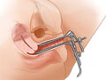
During the test, a dye (contrast material) is put through a thin tube. That tube is put through the vagina and into the uterus. Because the uterus and the fallopian tubes are hooked together, the dye will flow into the fallopian tubes. Pictures are taken using a steady beam of X-ray (fluoroscopy) as the dye passes through the uterus and fallopian tubes. The pictures can show problems such as an injury or abnormal structure of the uterus or fallopian tubes. They can also show a blockage that would prevent an egg moving through a fallopian tube to the uterus. A blockage also could prevent sperm from moving into a fallopian tube and joining (fertilizing) an egg. The test also may find problems on the inside of the uterus that prevent a fertilized egg from attaching (implanting) to the uterine wall.
Why It Is Done
A hysterosalpingogram (HSG) is done to:
- Check for a blocked fallopian tube. The test often is done for a woman who is having a hard time getting pregnant. An infection may cause severe scarring of the fallopian tubes and block the tubes. This can prevent pregnancy. Once in a while, the dye used during the HSG will push through and open a blocked tube.
- Find problems in the uterus, such as an abnormal shape or structure. The test can also look for an injury, polyps, fibroids, adhesions, or a foreign object in the uterus. These types of problems may cause painful menstrual periods or repeated miscarriages.
- See if tubal implants for permanent birth control are blocking the fallopian tubes.
- See if surgery to reverse a tubal ligation has been successful.
How To Prepare
Before a hysterosalpingogram (HSG), tell your doctor if you:
- Are or might be pregnant.
- Have a pelvic infection (pelvic inflammatory disease) or a sexually transmitted infection, such as gonorrhea or chlamydia.
- Are allergic to the iodine dye used or any other substance that has iodine. Also tell your doctor if you have asthma or are allergic to any medicines. Tell him or her if you have had a serious allergic reaction (anaphylaxis) from any substance. (For example, have you had a reaction to the venom from a bee sting or from eating shellfish?)
- Take a blood thinner, or if you have had bleeding problems.
- Have a history of kidney problems or diabetes, especially if you take metformin (such as Glucophage) to control your diabetes. The dye used during the test can cause kidney damage in people with poor kidney function. If you have a history of kidney problems, blood tests (creatinine, blood urea nitrogen) may be done before the test. These check to see that your kidneys are working well.
This test should be done 2 to 5 days after your menstrual period has ended. It should also be done before you ovulate the next month (unless you are using contraception). This is to avoid using X-rays during an early pregnancy. You may want to bring along a sanitary pad to wear after the test. That’s because some leakage of the X-ray dye may occur along with slight bleeding.
You may be asked to sign a consent form that says you understand the risks of the test and agree to have it done.
Talk to your doctor about any concerns you have about the need for the test, its risks, how it will be done, or what the results will mean. To help you understand the importance of this test, fill out the medical test information form( What is a PDF document? ).
How It Is Done
A hysterosalpingogram usually is done by a radiologist in the X-ray room of a hospital or clinic. A radiology technologist and a nurse may help the doctor. A gynecologist or a doctor who specializes in infertility (reproductive endocrinologist) also may help with the test.
Before the test begins, you may get a sedative to help you relax. You may also get ibuprofen to help relax your uterus so it will not cramp during the test. You will need to take off your clothes below the waist and drape a gown around your waist. You will empty your bladder. Then you will lie on your back on an exam table. Your feet will be raised and supported by stirrups. This allows your doctor to look at your genital area.
Your doctor will put a smooth, curved tool called a speculum into your vagina. The speculum gently spreads apart the vaginal walls. This allows the doctor to see the inside of the vagina and the cervix. The cervix may be held in place with a clamp called a tenaculum. The cervix is washed with a special soap. A stiff tube (cannula) or a flexible tube (catheter) is put through the cervix into the uterus. The X-ray dye is put through the tube. If the fallopian tubes are open, the dye will flow through them. It will then spill into the belly where it will be absorbed by the body. If a fallopian tube is blocked, the dye will not pass through. The X-ray pictures are shown on a TV screen during the test. If another view is needed, the exam table may be tilted or you may be asked to change position.
After the test, the cannula or catheter and the speculum are removed. This test usually takes 15 to 30 minutes.
How It Feels
You will most likely feel some cramping like menstrual cramps during the test. The amount of pain you have depends on what problems the doctor finds and treats during the test.
Risks
There is always a small chance of damage to cells or tissue from being exposed to any radiation. This can include the low levels of radiation used for this test. The chance of damage from the X-rays is generally very low compared with the possible benefits of the test.
There is a small chance of a pelvic infection after the test. Examples of these are endometritis and salpingitis. The chance may be higher for women who have had pelvic infections before. Your doctor may give you antibiotics if he or she thinks you might get a pelvic infection.
There is a small chance of damaging or puncturing the uterus or fallopian tubes during the test.
There is a small chance of an allergic reaction to the iodine X-ray dye. This is more common if you are allergic to any shellfish.
In rare cases, if an oil-based dye is used, the oil can leak into the blood. This can cause blockage of blood flow to a section of the lung (pulmonary embolism). But in most cases, this test uses water-based dyes.
After the test
After the test, some of the dye will leak out of the vagina. You also may have some vaginal bleeding for several days after the test. Call your doctor right away if you have:
- Heavy vaginal bleeding. (This means soaking more than one tampon or pad in 1 hour.)
- A fever.
- Severe belly pain.
- Vaginal bleeding that lasts for more than 3 to 4 days.
Results
A hysterosalpingogram (HSG) is an X-ray test. It looks at the inside of the uterus and fallopian tubes and the area around them.
|
Normal: |
The shape of the uterus and fallopian tubes are normal. The fallopian tubes are not scarred or damaged. The dye flows freely from the uterus and through the fallopian tubes and then spills normally into the belly. |
|
No objects (such as an intrauterine device, or IUD), tumors, or growths are seen in the uterus. |
|
|
Abnormal: |
Fallopian tubes may be scarred, malformed, or blocked so that the dye does not flow through the tubes and spill into the belly. Blocked fallopian tubes may be caused by pelvic inflammatory disease (PID) or endometriosis. |
|
The dye may leak through the wall of the uterus, showing a tear or hole in the uterus. |
|
|
An abnormal uterus may show tissue (called a septum) that divides the uterus. |
|
What Affects the Test
You may not be able to have the test, or the results may not be helpful, if:
- Your fallopian tube has a spasm. This may make a normal fallopian tube look blocked.
- The doctor can’t put a catheter in the uterus.
This test is not done on women who are having their period, are pregnant, or have a pelvic infection.
What To Think About
- In some cases, a pelvic ultrasound test may be done instead of a hysterosalpingogram (HSG) to find foreign objects in the uterus, such as an intrauterine device (IUD). To learn more, see the topic Pelvic Ultrasound.
- Some early tests to find the cause of infertility may include tests such as semen analysis and blood tests for luteinizing hormone (LH), progesterone, or follicle-stimulating hormone (FSH). If these tests can’t find the cause of infertility, an HSG may be done. To learn more, see the topic Infertility Testing.
- An HSG is done mainly for women who are having a hard time getting pregnant. Some studies show that this test may help a woman’s chance of becoming pregnant. That is because the dye may remove the mucus plugs, straighten the fallopian tubes, and break through thin scar tissue.
- A hysteroscopy may be done instead to look at the uterus. A test called a laparoscopy may also be done instead to look at the fallopian tubes. A laparoscopy does not show whether the fallopian tubes are open, unless dye is injected during the test.
- Another test, called a sonohysterogram (SHG), may be more accurate for looking at uterine fibroids or polyps. SHG uses ultrasound to watch the movement of a salt solution (saline) that is injected into the uterus. SHG does not use X-rays or an iodine dye.
- If a blocked fallopian tube is the cause of infertility, an oil-based dye may be used during an HSG to remove the blockage. Some studies show that an oil-based dye may open up a blockage better than a water-based dye. Other studies have shown no difference between the two dyes.
- Be sure your doctor knows if you take metformin (such as Glucophage) for diabetes or for any other reason, such as polycystic ovary syndrome (PCOS). This is important because of the possible interaction with the dye used in this test.
Current as of: February 19, 2019
Author: Healthwise Staff
Medical Review:Sarah A. Marshall, MD – Family Medicine & Kathleen Romito, MD – Family Medicine & Martin J. Gabica, MD – Family Medicine & E. Gregory Thompson, MD – Internal Medicine & Deborah A. Penava, MD, FRCSC, MPH – Obstetrics and Gynecology


This information does not replace the advice of a doctor. Healthwise, Incorporated, disclaims any warranty or liability for your use of this information. Your use of this information means that you agree to the Terms of Use. Learn how we develop our content.