Electroencephalogram (EEG)
Test Overview
An electroencephalogram (EEG) is a test that measures and records the electrical activity of your brain. Special sensors called electrodes are attached to your head. They’re hooked by wires to a computer. The computer records your brain’s electrical activity on the screen. Or it may record the activity on paper as wavy lines. Changes from the normal pattern of electrical activity can show certain conditions, such as seizures.
Why It Is Done
An EEG may be done to:
- Check for epilepsy and see what type of seizures are occurring. EEG is the most useful and important test for checking if someone has epilepsy.
- Check for problems with loss of consciousness or dementia.
- Help find out a person’s chance of getting better after a change in consciousness.
- Find out if a person who is in a coma is brain-dead.
- Study sleep disorders, such as narcolepsy.
- Watch brain activity while a person is getting general anesthesia for brain surgery.
- Help find out if a person has a physical problem or a mental health problem. Physical problems include problems in the brain, spinal cord, or nervous system.
How To Prepare
Before the day of the EEG test, tell your doctor if you are taking any medicines. Your doctor may ask you to stop taking certain medicines before the test. They include sedatives and tranquilizers, muscle relaxants, sleeping aids, and medicines used to treat seizures. These medicines can affect your brain’s usual electrical activity. Taking them may affect your test results.
Do not eat or drink things that have caffeine for 12 hours before the test. This includes coffee, tea, cola, and chocolate.
The electrodes will be attached to your scalp. Make sure that your hair is clean before the test. Don’t put sprays, oils, creams, or lotions in your hair. Shampoo your hair and rinse with clear water the night before or the morning of the test. Do not put any hair conditioner or oil in your hair after you wash it.
To find certain types of abnormal electrical activity in the brain, you may have to be asleep during the test. You may be asked not to sleep at all the night before the test. Or you may need to sleep less (about 4 or 5 hours) by going to bed late and getting up early. If your child is going to be tested, try to keep him or her from taking naps just before the test. If you know that you are going to have an EEG with little or no sleep, plan to have someone drive you to and from the test.
How It Is Done
An EEG may be done in a hospital or in a doctor’s office. An EEG technologist does the test. The EEG record is read by a doctor who is trained to diagnose and treat problems that affect the nervous system (neurologist).
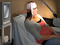
You will be asked to lie on your back on a bed or table. Or you may sit in a chair with your eyes closed. The EEG technologist will attach several flat metal discs (electrodes) to different places on your head. A sticky paste is used to hold them in place. Instead of separate electrodes, you may wear a cap with several fixed electrodes. In rare cases, the electrodes may be attached to the scalp with tiny needles.
The electrodes are hooked by wires to a computer that records the electrical activity in the brain. A machine can show the activity as a series of wavy lines on a piece of paper. Or the activity may be shown as an image on the computer screen.
You will need to lie still with your eyes closed during the recording. The technologist will watch you directly or through a window during the test. The recording may be stopped from time to time. This allows you to stretch and change your position.
The technologist may ask you to do different things during the test to see what activity your brain does at that time.
- You may be asked to take deep and rapid breaths (hyperventilate). Usually you will take 20 breaths a minute for 3 minutes.
- You may be asked to look at a bright, flashing light called a strobe.
- You may be asked to go to sleep. If you can’t fall asleep, you may get a sedative to help you sleep. If an EEG is being done to check a sleep problem, your brain’s electrical activity may be recorded all night.
An EEG takes 1 to 2 hours. After the test, you may do your normal activities. But if you had little or no sleep or were given a sleep medicine, have someone drive you home after the test.
How It Feels
There is no pain during an EEG.
Paste may be used to hold the electrodes in place. Some of the paste may stick in your hair after the test. You will have to wash your hair to get it out. If needle electrodes are used (which is rare), you will feel a brief, sharp prick when each electrode is put in. It will feel kind of like having a hair pulled out. If electrodes are placed in your nose, they may tickle. Rarely, this may cause some soreness or a small amount of bleeding for 1 to 2 days after the test.
If you are asked to breathe fast, you may feel lightheaded or have some numbness in your fingers. This is normal. It will go away a few minutes after you start breathing normally again.
Risks
An EEG is a very safe test. The electrical activity of your brain is recorded. But no electrical current is put into your body. An EEG is not the same as electroshock (electroconvulsive) therapy.
If you have a seizure disorder such as epilepsy, the flashing lights may trigger a seizure. Or a seizure may happen if you hyperventilate. If it happens, the technologist is trained to take care of you during the seizure.
Results
An electroencephalogram (EEG) is a test that measures and records the electrical activity of your brain. Special sensors called electrodes are attached to your head. They’re hooked by wires to a computer. EEG test results are ready on the same day or the next day.
There are several types of brain waves.
- Alpha waves are present only when you’re awake with your eyes closed but you are mentally alert. Alpha waves go away when your eyes are open or you are concentrating.
- Beta waves are normally found when you are alert or have taken high doses of certain medicines, such as benzodiazepines.
- Delta waves are normally found only in young children and in people who are asleep.
- Theta waves are normally found only in young children and in people who are asleep.
|
Normal: |
In adults who are awake, the EEG shows mostly alpha waves and beta waves. |
|
The two sides of the brain show similar patterns of electrical activity. |
|
|
There are no abnormal bursts of electrical activity and no slow brain waves on the EEG tracing. |
|
|
If flashing lights are used during the test, one area of the brain (the occipital region) may have a brief response after each flash of light. But the brain waves are normal. |
|
|
Abnormal: |
The two sides of the brain show different patterns of electrical activity. This may mean that there’s a problem in one area or side of the brain. |
|
The EEG shows sudden bursts of electrical activity called spikes. Or the test shows sudden slowing of brain waves in the brain. These changes may be caused by a brain tumor, infection, injury, stroke, or epilepsy. When a person has epilepsy, the location and exact pattern of the abnormal brain waves may help show the type of epilepsy or seizures. In many people with epilepsy, the EEG may appear normal between seizures. An EEG by itself does not diagnose or rule out epilepsy or a seizure problem. |
|
|
The EEG records changes in the brain waves that may not be in just one area of the brain. A problem that affects the whole brain may cause these kinds of changes. This includes drug intoxication, infections (encephalitis), and metabolic disorders (such as diabetic ketoacidosis). These problems change the chemical balance in the body, including the brain. |
|
|
The EEG shows delta waves or too many theta waves in adults who are awake. This may mean that there is a brain injury or brain illness. Some medicines can also cause this. |
|
|
The EEG shows no electrical activity in the brain. This is a “flat” or “straight-line” EEG. This means that brain function has stopped. It’s usually caused by lack of oxygen or blood flow inside the brain. It may happen when a person has been in a coma. In some cases, severe sedation from drugs can cause a flat EEG. |
What Affects the Test
You may not be able to have the test, or the results may not be helpful, if:
- You move too much.
- You take certain medicines. This includes medicines used to treat seizures (antiepileptic medicines), sedatives, tranquilizers, and barbiturates.
- You drank coffee, soda, or tea, or you ate other foods that have caffeine before the test.
- You are unconscious from severe drug poisoning or a very low body temperature (hypothermia).
- Your hair is dirty, oily, or covered with hair spray or other hair products. This can cause a problem with how the electrodes are placed.
What To Think About
- If the doctor thinks that a person has epilepsy but the EEG is normal, the EEG technologist may have the person look at a flashing light. Or the person may be asked to breathe fast and deep (hyperventilate) or sleep during the test. These techniques sometimes show epileptic EEG patterns that did not show up at first. If epilepsy is suspected after the first EEG, the doctor may repeat the EEG more than once.
- An EEG done during a seizure will almost always show electrical patterns that aren’t normal. This makes an EEG useful when a doctor thinks that a person is having psychogenic nonepileptic seizures. These are also called pseudoseizures. They have no physical cause. But they can be caused by stress, emotional trauma, or mental illness. These seizures do not cause abnormal electrical activity in the brain. They will not show abnormal EEG results.
- There could still be a problem in the brain even when the EEG is normal.
- Other tests that may also be done include:
- Video EEG. Video EEG records seizures on video and on a computer. This way the doctor can see what happens just before, during, and right after a seizure. This test can help find the specific area of the brain that the seizures may be coming from. It also helps in diagnosing psychogenic seizures, which may look like real seizures but do not affect the electrical activity in the brain. Video EEG may be used short-term or long-term.
- Short-term monitoring is done on an outpatient basis. It may last up to 6 hours.
- Long-term monitoring is done in the hospital. It may last 3 to 7 days.
- Brain mapping. This is a fairly new method that is very similar to EEG. Electrodes are placed on the person’s scalp to transmit the brain’s electrical activity. Then a computer makes a color-coded map of signals from the brain. It is sometimes done to find a specific problem area in the brain that has already shown up on a regular EEG. Doctors are still not sure how brain mapping could be best used.
- Ambulatory EEG monitoring. During this test, the person is able to move around. The test allows for electrical activity in the brain to be recorded for a long time. Fewer electrodes are attached to the person. The person carries a small, portable recording unit. The test may last for a full day or more. The person can leave the hospital. Ambulatory EEG monitoring is not as accurate as a regular EEG.
- Video EEG. Video EEG records seizures on video and on a computer. This way the doctor can see what happens just before, during, and right after a seizure. This test can help find the specific area of the brain that the seizures may be coming from. It also helps in diagnosing psychogenic seizures, which may look like real seizures but do not affect the electrical activity in the brain. Video EEG may be used short-term or long-term.
Current as of: March 28, 2019
Author: Healthwise Staff
Medical Review:Anne C. Poinier, MD – Internal Medicine & E. Gregory Thompson, MD – Internal Medicine & Adam Husney, MD – Family Medicine & Martin J. Gabica, MD – Family Medicine & Karin M. Lindholm, DO – Neurology
This information does not replace the advice of a doctor. Healthwise, Incorporated, disclaims any warranty or liability for your use of this information. Your use of this information means that you agree to the Terms of Use. Learn how we develop our content.